


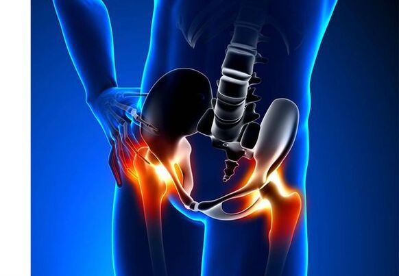
Osteoarthritis of the hip joint (coxarthrosis)- This is a chronic degenerative joint disease that leads to the deformation of bone tissue.In the case of coksart, all components of the joint are involved in the pathological process: articular cartilage, bone structures in addition to cartilage, synovial bowl, ligaments, capsule and adjacent muscles.In diseases, articular cartilage is destroyed, micro reductions of bones and osteophytes (bone growth) occur, and inflammation of the muscle league apparatus of the hip joint occurs.
In the world, every fifth person complains about common problems with joints.This can be both pain and a restriction of movement in the joints and a combination of these symptoms.Every second outpatient view falls on patients with bone muscular disorders, while 66 % of cases are people under the age of 65.According to the latest epidemiological research, the prevalence of arthrosis of the knee and hip joint in the adult population is 13 %.
Risk factors for the development of coxarthrosis:
- Genetic predisposition.A common cause of coksart rose of the hip joints is the innate or acquired mutation of the type of type -ii prollages.
- Older age.The likely cause of the prevalence of osteoarthritis in old age is a discrepancy between the harmful effect on the common cartilage of the external environment and its restoring skills.
- Floor.Women suffer from arthrosis more often than men.This is due to the effects of the influence of female sex hormones from estrogen to the bone mineral metabolism.However, the influence of the soil is ambiguous - in the opinion of some authors, in contrast to damage to other joints, there are no differences in the sexual basis for coksart rose: in men, the osteoarthritis of the hip joint can be found as often as in women.
- Excess body weight.The relationship is proven between the excess body mass and the occurrence of osteoarthritis.Excess adhesive tissue increases the harmful load on the cartilage.In addition, fatty tissue generates pro -attachment enzymes that damage the cartilage tissue.
- Frequent development of bones and joints.In accordance with studies, 80 % of coxarthrosis, which occurs for no obvious reason, is associated with defects that have not previously been diagnosed in the development of dysplasia and subluxation of the hip joints.
- Heavy physical work.An excess strain on the hip joints with certain types of physical contractions can lead to damage to the joints and the formation of osteoarthritis.Agriculture, excavators and people of similar working specialties are at risk.
- Injuries.The risk of coxarthrosis increases after a violation of the hip joint.In addition, both an injured joint and both can be involved in the process.
- Professional sport.Professional sport can cause the occurrence of coxarthrosis both due to the excessive stress on the joints and due to injuries.The potentially dangerous sports include heavy athletics, athletics jump and parachut.
- Bones and common diseases.
- Endocrine pathologies- Hypothyroidism, hypoparathyroidism, acromegaly (impaired function of the front pituitary), diabetes, obesity.
If similar symptoms are found, consult a doctor.Do not do yourself - it is dangerous for your health!
Symptoms of arthrosis of the hip joints
The main symptoms of coxarthrosis are: pain, mobility restrictions and crunch in the joints, their deformation, functional shortening of the lower extremity and periodic swelling in the joints.
Pain of different intensity.The pain in the joint is initially insignificant and arise for a short time.They appear or intensify when walking or with other physical efforts, for example in squats, tendencies and lifting weight.While the disease develops, pain increases and even a long break does not bring relief.In addition, pain occurs in a position with longer immobility and fixation of the joint.
The patients complain about the so -called "start" scales in the hip lines after bed, in a car and another longer immobility."Pain for coxarthrosis" does not take longer than 30 minutes.The pain increases during hypothermia or in a stressful situation.They can be localized in the area of the buttocks or the groin area on the front or side surface of the thigh.With the spread of pain over the nerve of the lumbar plexus, it can be transferred to the thighs removed from the middle of the body or in the knee.Sometimes the pain applies to the lumbosacral spine and the coccyx.
Restriction of common mobility.Movements in the hip joint with coksart rose are limited due to pain.At the same time, the rotation (both inside and outside) and the lower extremity (movement in the middle of the body) is more often disturbed, but can be limited (movement from the middle body axis) as well as flexion and extension.The inability to make passive movements in the joint due to a pronounced pain syndrome causes a compensatory pelvic distortion.The patient's walk changes, the buttocks stretch back, the body dodges forward when it transfers weight to the damaged side.In the case of bilateral damage in patients with coksart, a "duck passage" is formed.
With regular occurrence of coxarthrosis occurs regularlySwelling in the jointwhich can be invisible due to the muscle and fat layer.The disease is also characteristicCrystal in the joints during movement, its gradual deformation and functional shortening of the lower limb.
A joint is often affected by the disease, then the process applies to others.Sometimes osteoarthritis influences several joints at the same time and polyosostoarthritis occurs.Polyosteoarthrosis is characteristic of older people or with hereditary disposition and at the same time diseases - diseases of bones, joints and endocrine diseases.
Pathogenesis of the arthrosis of the hip joints
With the pathogenesis of the arthrosis of the hip joints, an important role is played by mechanical damage (injuries and microtraumas due to increased physical exertion on the joint) and genetic, hormonal and metabolic factors.It is often not possible to find out which factor has influenced the development of the disease in a certain patient, but often the disease develops after tissue damage in the event of mechanical injury.
Damage to the tissue stimulates the division of cartilage tissue cells (chondrocytes), while the production of pro -inflammatory cytokines increases, which are normally available in cartilage.The cytokines start the inflammatory process, for example under the influence of anti-inflammatory cytokin il-1, enzymes that destroy the cartilage of the joint.Under the influence of cytokines, the production of the TSOG 21 enzyme and other substances that have a toxic influence on the cartilage also increases.
Synovites also play a major role in the development of coxarthrosis - inflammatory diseases of the synovial shell of joints or ligaments with the accumulation of liquid in the cavity.
A decrease in the elasticity and strength of the joint cartilage associated with the metabolic disorders leads to a decrease in resistance to mechanical stress.In the case of coksart, all components of the joints are involved in the pathological process, including a subchondral bone.Due to the fact that large joints of the lower extremities make up large joints of the body, they have a significant mechanical voltage, since micro -forms occur in the subchondral plate and the cartilage.As a result of microvelomas, the subchondral bone is compressed, which leads to regional growth of bone tissue - osteophytes.And this in turn stimulates the further deterioration of the articular cartilage.
In some cases, the arthrosis of the hip joint is inherited.Hereditary osteoarthritis is supposedly a polygenic heritage - due to the effect of many genes, each of which is weakly influenced.The cause of some diseases is a mutation in genes that encodes macromolecules made of articular cartilage, causing its fractures.Genes who are responsible for the division of chondrocytes can also suffer.In addition, metabolic disorders are inherited, such as pyrophosphate arthropathy - a disease in which crystals from calcium pyrophosphate accumulate in the articular cartilage and synovial fluid.
Classification and stages of the development of the arthrosis of the hip joints
Depending on the causes of the disease, the coxarthrosis is divided into two main forms: primarily (idiopathic) and secondary (from or due to other diseases).
Primary Coksartrose:
- Located (only have a hip joints):
- one -sided;
- bilateral.
- Generalized (polyosteoarthrosis) with a lesion of at least three joint groups (e.g. hips, knees and small joints of brushes or feet).
Secondary arthrosis:
- Post -Traumatic:
- Acute - as a result of an acute injury;
- Chronic - due to physical education or due to professional activities.
- Metabolic diseases (occonosis, hemochromatosis, Wilson disease, gaucher disease).
- Congenital pathologies and development defects (congenital dysplasia of the hip joint, pertes disease, slipping the epiphysis of the femur, hypermobility syndrome, shortening of the lower limbs, scoliosis, bone dysplasia).
- Endocrine pathologies (acromegaly, hypothyroidism, diabetes mellitus, hyperparathyroidism, obesity).
- Calcium salt (pyrophosphate arthropathy, calcification of tendonitis).
- Bone and joint diseases (rheumatoid arthritis, psoriasis arthritis, pedestically disease, avascular necrosis, infections).
According to clinical manifestations, the following forms of coxarthrosis are differentiated:
- Not very symptomic.
- Manifested, manifested by bright clinical symptoms:
- quickly progressively, in which the symptoms develop in the first four years from the beginning of the disease;
- Slowly progressive - clinically significant symptoms occur after five years of the disease.
In accordance with the X -Ray image, two types of osteoarthritis of the hip joints can be identified:
- Hypertrophic - with signs of an increased reparative reaction (lesions are replaced by a new tissue, for example osteophytes appear);
- Atrophic (acceptance of the tissue volume).
The stages of the disease can be determined radiologically and clinically.In order to determine the radiological stage of the arthrosis of the hip joint, the classification of Kellgren and Lawrence (1957) is most frequently used.
Arthrosis stadiums in radiological classification
| stage | Sign |
|---|---|
| 0 | There are no signs of osteoarthritis in X -Ray pictures |
| 1 | The common gap is not changed, individual regional osteophytes are made visible |
| 2 | The common gap is not changed, significant regional osteophytes are made visible |
| 3 | The amount of the common gap is moderately reduced, significant regional osteophytes are made visible |
| 4 | The height of the joint column is significantly reduced, significant regional osteophytes and subchondral osteosclerosis are made visible (bone tissue compression under the lower surface of the cartilage with the structure of the cartilage) |
Classification (1961) is used to determine the clinical stage of the disease that uses both clinical signs and visualization criteria.
Clinical stages of arthrosis
| stage | Sign |
|---|---|
| 0 | The joint gap is clear and unevenly narrowed, the edges of the joint crawl are slightly pointed (initial osteophytes), a slight restriction of the movements is determined |
| 1 | The joint gap is significantly narrowed (50-60 %), significant osteophytes, subchondral osteocosclerosis and cystic enlightenment in bone epiphysees;The clinic is dominated by restricting mobility in the joints, a rough crunch during movements, insignificant or moderate muscle atrophy |
| 2 | Deformation, stiffness of the joint;The joint column is narrowed by more than 60-70 % of the standard or not missing, extensive osteophytes, subchondral cysts and articular "mice" are visualized in visualized bones, cartilage or mixed pathological formations in the joint cavity |
Complications of the arthrosis of the hip joints
In the case of coxarthrosis, all complications are connected with pathological changes in the joints.
The course of the coksart rose can be complicated by local inflammatory processes:
- Bursit - inflammation of synovial bags in the joints;
- Tendovaginitis - inflammation of the inner shell of the vagina of muscle tendons;
- Tunnel syndrome pinches of the nerve due to the formation of large osteophytes or with joint deformation.
With the progress of coxarthrosis and the transition to clinical stages II and III, the pain limits the mobility of the joint, and over time the joint core (fibrous, bone or cartilage) occurs, accompanied by its complete immobility.
A significant common deformation can lead to thisFractures or aseptic necrosis of bones.For coksart rose, aseptic necrosis of the thigh head is the most impressive complication.
With pronounced coksart rose, it can occurSubbluxation and transfer of the jointas well as the penetration of the thigh head into the pelvic cavity.The transfers and subluxation of the hip joint lead to pain (initially acute, then dull and painful), during walking and other physical exertion as well as a deformation of the joint, lame and sometimes to shorten the affected member.
Despite the lack of systemic manifestations of arthrosis, even in modern clinical practice, the associated diseases are given more attention.These are pathological conditions that exist or arise against the background of the current illness.In connection with inflammatory reactions that arise during osteoarthritisCardiovascular diseases.A decrease in physical activity due to pain and restricting joint mobility leadsObesity, depression and deterioration in quality of life.With longer use of non -steroidal anti -inflammatory drugs,The upper stomach intestine cuts are affected,And alsoThe risk of cardiovascular pathologies and kidney diseases increases.
Diagnosis of the arthrosis of the hip joints
The diagnosis of "Coksartrose" is based on clinical manifestations and radiological examination.There are no characteristic laboratory signs for diagnosis of osteoarthritis.
Among the clinical manifestationsThe main diagnosis of arthrosis of the hip joint is the pain and its character.Pain in the arthrosis of the hip joint gradually occurs over several years (sometimes several months with a rapidly progressive form).The pain occurs during physical exertion or in a standing position or improves.If the patient feels pain alone, inflammation (synovitis) has connected.The statement is found up to 30 minutes in the morning and with longer immobility.
The restriction of common mobility is gradually increasing, this applies to both active and passive movements.The joints are deformed in the development of the disease, and functional shortening of the limb length can occur.
During a physical examinationThere is a restriction of joint mobility, its deformation, the shortening of the limbs, the pain when palpating the joint and a large turn of the femoral muscle atrophy.
Laboratory methodsIt is not necessary for the diagnosis of osteoarthritis of the hip joints.However, they can be used for the differential diagnosis of coxarthrosis with arthritis (rheumatoid and chronic), since no inflammatory changes in the total blood test and rheumatoid factor and uric acid level are not increased in arthrosis.In addition, contraindications for drug treatment methods are uncovered in laboratory tests.
Instrumental methodsFor the diagnosis of arthrosis of the hip joints:
- Radiography- This is the main method for diagnosing osteoarthritis of the hip joints.The X -ray image determines the changes that are characteristic of CoksartRose: narrowing of the common gap, osteophytes, erosion and ulcers of the cartilage, the subchondral cysts and osteosclerosis.The X -Ray examination is a classic method for diagnosing coxarthrosis, and radiological signs on the basis of the classification of coxarthrosis.However, other visualization methods of the connection are currently being used, such as:B. ultrasound and magnetic resonance imaging.
- Ultrasound examination (ultrasound) -The advantage of ultrasound is in the absence of a radial stress on the body.
- Magnetic resonance imaging (MRI)- Compared to other methods, you can visualize the joint damage more clearly.
- Arthroscopy- -Allows you to identify damage to the articular cartilage: from the chondromation zones (softening the articular cartilage) with a diameter of less than 10 mm to deep cracks that penetrate to the subchondral bone and the formation of deep ulcers.Superficial and medium cracks and surface erosion can also be made visible.
The identification of Coksartrose usually shows no special difficulties, but when assessing a specific clinical situation, the possible secondary origin of the arthrosis of the hip joints must remember (as complications of other diseases, for example with endocrine diseases).
Treatment of the arthrosis of the hip joints
The treatment of the arthrosis of the hip joints can be both conservative (medication and not united) or operational.Conservative treatment is used in 1-2 stages of the disease, surgical levels of 3 stadiums.Surgical treatment can be recommended in 2 stages with persistent pain and a lack of reaction to conservative therapy.
The goals of conservative therapy:
- Improvement of quality of life - reduce pain and increase joint mobility;
- Stop or slow down the development of the disease.
The non -drug treatment methods include:
- Unloading the hip joint (acceptance of body weight, the creation of additional support and the transfer of body weight to stick or crutch);
- Physiotherapy sports lessons;
- Physiotherapeutic treatment methods.
The treatment of coxarthrosis begins with non -displaced methods.An important role is taken on physiotherapy exercises.In the event of severe pain, the patient should use the support.With a pronounced illness and the presence of contraindications for endoprosthetics, support for life must be used.
Healing therapy for cuxartrosisIncludes medication that reduces the symptoms of the disease.These are analgesics and medicines from the group of non -steroidal anti -inflammatory drugs (NSAIDS).The NSAIDs are divided into non -election and selectively.
Analgetics and NSAIDS for the arthrosis of the hip joint are used for a short time to relieve pain and inflammation.At the moment there is no proven advantage of a non -steroidal anti -inflammatory agent compared to another, so the choice of a certain drug depends on the side effects and a specific clinical situation.
It must be remembered that NSAIDS have a number of side effects.When taking it, the mucous membrane of the stomach and duodenum is affected, with ulcers and bleeding possible.A number of NSAIDS has toxic to the liver and kidneys.In addition, NSAIDS disturb the thrombocyte aggregation, and as a result, the patient is disturbed by thrombosis and there is a tendency towards bleeding.The NSAIDS with longer use suppress the processes of hematopoies and can cause aplastic anemia and agranulocytosis.The reception of selective NSAIDs leads to considerable complications.
Ointments and gels that are used locally cause fewer side effects than oral products.Medicines with heating and reducing pain are used to treat osteoarthritis.You can contain turpentine, menthol, nicotine acid ester, salicylate and beeergift.The NSAIDs also have a good effect.
In the absence of the effects of analgesic and NSAIDS or if it is impossible to choose the optimal dose of the drug, pain relievers of the central effect can be prescribed at short notice.
In inflammation, the intra -karticular administration of corticosteroids is used.Corticosteroids are not used more than 2-3 times a year because more frequent use can lead to cartilage degeneration.
Slowly effective medication weaken the symptoms of the disease, including chondroprotectors, inappropriate connections from avocados or soy, hyaluronic acid.These drugs are included in the recommendations of the European anti -Rematic league to treat the arthrosis of the hip joints.Preparations reduce the pain and improve joint mobility.
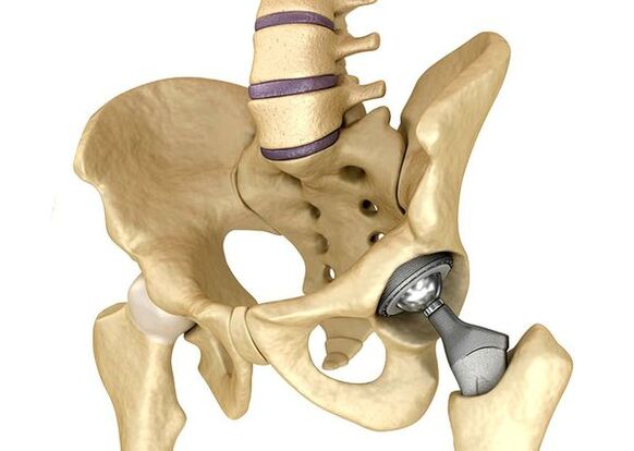
Endoprosthetics of the hip jointsStage III is used in severe cases if pain syndrome cannot be eliminated and the mobility of the joint is significantly limited.The prosthetics of the hip joint leads to a reduction in pain syndrome, an improvement in the functional state of the joint and the quality of the patient's life.The effect remains for 10-15 years that a second operation may be required.During the operation, the hip joint is replaced by artificial imitation of ceramics, metal (most frequently used titanium prostheses) or polymer.
Forecast.prevention
The forecast of arthrosis of the hip joints in terms of the patient's life is favorable, but the disease often leads to disabilities.According to the World Health Organization, 80 % of the elderly patients with coxarthrosis have a violation of mobility, and 25 % cannot do everyday affairs.In this regard, the primary prevention of arthrosis of the hip joints is important.
Prevention measures:
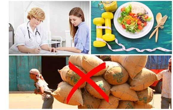
- Reduce body weight.It is necessary to adapt the diet to reduce the weight and load of the joint.In addition, a decrease in the volume of the adipose tissue reduces the amount of inflammatory mediators that have released it.
- Avoid severe physical work and sports overloads.Physical overloads are often the cause of osteoarthritis of the hip joints, while moderate physical activity, on the contrary, improves the condition of the articular cartilage, maintains its normal mobility and reduces the load on other joints.
- Correct the underlying disease.If the patient is detected in diseases that can lead to secondary coksart rose (endocrine, rheumatic and others), the underlying disease is required.The normalization of the hormonal background and the achievement of the continued remission of rheumatic diseases are both the primary prevention of osteoarthritis and it enables it to slow down.
- Lead a healthy lifestyle.A balanced diet with a sufficient content of herbal and animal protein, polyunsaturated fatty acids and the limitation of simple carbohydrates as well as moderate physical activity avoid the occurrence of coxarthrosis in the presence of risk factors.
The prevention of diseases of the hip joint in neonatology and pediatrics is currently mandatory.Over time, the adapted innate dysplasia of the hip joint reduces the risk of coxarthrosis in adulthood.